

© All rights reserved galwayclinic.com 2013


Galway Clinic Facebook page
Tel: +353 (0) 91720170 brendan.ocochlain@galwayclinic.com
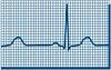
Electrocardiogram (ECG ) :
A recording of the electrical impulses traveling through the heart muscle. An ECG is recorded on graph paper, through the use of electrodes (small, sticky patches) that are attached to your skin on the chest, arms and legs.
Echocardiogram
A type of ultrasound used to provide a view of the heart to determine if there is heart muscle or valve disease that may be causing an arrhythmia. This test may be performed at rest or with activity. During an echo test, ultrasound (high-frequency sound waves) from a hand-held wand placed on your chest provides pictures of the heart's valves and chambers and helps the sonographer evaluate the pumping action of the heart. Echo is often combined with Doppler ultrasound and colour Doppler to evaluate blood flow across the heart's valves.Why is an echocardiogram performed?
The test is used to: • Assess the overall function of your heart • Determine the presence of many types of heart disease, such as valve disease, myocardial disease, pericardial disease, infective endocarditis, cardiac masses and congenital heart disease • Follow the progress of valve disease over time • Evaluate the effectiveness of your medical or surgical treatmentsWhat happens during the test?
You will be given a hospital gown to wear. You’ll be asked to remove your clothing from the waist up.


Galway Site Design

Galway Clinic Facebook page
Electrocardiogram (ECG ) :
A recording of the electrical impulses traveling through the heart muscle. An ECG is recorded on graph paper, through the use of electrodes (small, sticky patches) that are attached to your skin on the chest, arms and legs.
Echocardiogram
A type of ultrasound used to provide a view of the heart to determine if there is heart muscle or valve disease that may be causing an arrhythmia. This test may be performed at rest or with activity. During an echo test, ultrasound (high-frequency sound waves) from a hand- held wand placed on your chest provides pictures of the heart's valves and chambers and helps the sonographer evaluate the pumping action of the heart. Echo is often combined with Doppler ultrasound and colour Doppler to evaluate blood flow across the heart's valves.Why is an echocardiogram performed?
The test is used to: • Assess the overall function of your heart • Determine the presence of many types of heart disease, such as valve disease, myocardial disease, pericardial disease, infective endocarditis, cardiac masses and congenital heart disease • Follow the progress of valve disease over time • Evaluate the effectiveness of your medical or surgical treatmentsWhat happens during the test?
You will be given a hospital gown to wear. You’ll be asked to remove your clothing from the waist up.